
Cell Line Profile Nb2-11
(ECACC catalogue no. 97041101)
Cell line history
Nb2-11 is a clone of the Nb-2 rat lymphoma line deposited by Drs Gout and Friesen while at the Department of Cancer Endocrinology, British Columbia Cancer Agency, Vancouver. The cell line was originally developed from a transplant of a lymphoma which developed in the thymus/lymph node of a male Noble (Nb) strain rat following treatment with oestrogen. These cells originate from pre-T cells, and depend on mammalian lactogens, for example prolactin (Gout, Beer and Noble, 1980) to proliferate. Alternatively, the addition of IL-2 (interleukin-2) can also stimulate its growth (Cantrell and Smith, 1984). If Noble rats are injected with Nb2 or its clones, malignant tumours will start to develop. These tumours are highly sensitive to treatment with vinca alkaloids (Gout, Noble and Beer, 1986). The Noble strain rat and the Nb-2 cell line were subjected to karyotypic analysis, which found that the karyotype of the prolactin dependant Nb-2 cell lines had a number of defined chromosomal abnormalities compared to the rat strain which was developed (Horsman, Masui and Gout, 1991).
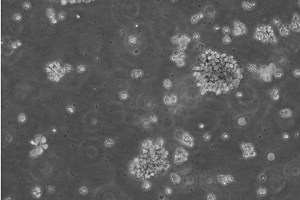
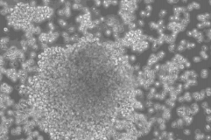
48 hours post resuscitation Prior to cryopreservation
Key characteristics
Nb2-11 cells are lymphoblastoid cells which grow in suspension and clump to form clusters when they are close to full confluence.
Applications
Nb2-11 cells have been used in a variety of applications – for example, they were used as part of a cell proliferation assay to assess the effect of exposing Nb2-11 cells to concentrations of hGH (a lactogenic hormone) (Nguyen et al, 2014). Another example of its application is an experiment which used ultrasonic sound waves to try and manipulate live Nb2-11 cells – that being their alignment, translocation and concentration (Siddique et al, 2014). Nb2-11 cells also present a model for tumour growth (Horsman, Masui and Gout, 1991), and putative anticancer treatments can be applied to the cells to assess their effectiveness. The cell line has also been used in a study to compare histochemical and immunochemical methods for the analysis of prolactin binding sites on Nb2-11 (Michel and Parsons, 1990).
Culture tips
Nb2-11 cells should be cultured in Fischer's medium (Life Technologies product number: 21475025) with the addition of 10% Foetal Bovine Serum (FBS), 10% Horse Serum (gelding), 0.075% Na Bicarbonate, 0.05mM 2-Mercaptoethanol (2ME) and 2mM Glutamine. At resuscitation, cultures should be passaged between 3-5x10⁵ cells/ml. Once established, cultures should be seeded at 4x10³ cells/ml for culture periods of 96 hours, and 1.2x10⁴ cells/ml for culture periods of 72 hours. The flasks should be kept at 37°C with 5% CO2, and checked daily. Do not allow the cell density to exceed 9x10⁵ cells/ml, as they will reach maximum confluence and will not continue to grow. The culture doubling time of these cells is approximately 12 hours.
Key References
-
Gout, P.W., Beer, C.T. and Noble, R.L., 1980. Prolactin-stimulated growth of cell cultures established from malignant Nb rat lymphomas. Cancer Research, 40(7), pp.2433-2436.
-
Cantrell, A. and Smith, K., 1984. The interleukin-2 T-cell system: a new cell growth model. Science, 224, pp. 1312.
-
Gout, P.W., Noble, R.L. and Beer, C.T., 1986. Cultured Nb rat lymphoma cells in endocrine and cancer research. Biochemistry and Cell Biology, 64(7), pp.659-666.
-
Nguyen, M.T., Koo, B.K., Vu, T.T.T., Song, J.A., Chong, S.H., Jeong, B., Ryu, H.B., Moh, S.H. and Choe, H., 2014. Prokaryotic soluble overexpression and purification of bioactive human growth hormone by fusion to thioredoxin, maltose binding protein, and protein disulfide isomerase. PLoS One, 9(3), p.e89038.
-
Siddique, A.K.M., Kim, C., Cho, S.H., Ahn, B. and Yoon, D.J., 2014. Use of frequency swept ultrasound to manipulate Nb2-11 live cells in a microfluidic channel. Journal of the Korean Physical Society, 64(2), pp.226-231.
-
Horsman, D.E., Masui, S. and Gout, P.W., 1991. Karyotypic changes associated with loss of prolactin dependency of rat Nb2 node lymphoma cell cultures. Cancer research, 51(1), pp.282-287.
-
Michel, E. and Parsons, J.A., 1990. Histochemical and immunocytochemical localization of prolactin receptors on Nb2 lymphoma cells: applications of confocal microscopy. Journal of Histochemistry & Cytochemistry, 38(7), pp.965-973.