
Cell line profile MDA-MB-231
(ECACC catalogue no. 92020424)
Cell line history
The MDA-MB-231 cell line is an epithelial, human breast cancer cell line that was established from a pleural effusion of a 51-year-old caucasian female with a metastatic mammary adenocarcinoma1 and is one of the most commonly used breast cancer cell lines in medical research laboratories.
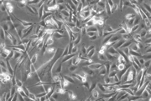
MDA-MB-231 cells in culture
Key characteristics
MDA-MB-231 is a highly aggressive, invasive and poorly differentiated triple-negative breast cancer (TNBC) cell line as it lacks oestrogen receptor (ER) and progesterone receptor (PR) expression, as well as HER2 (human epidermal growth factor receptor 2) amplification2,3. Similar to other invasive cancer cell lines, the invasiveness of the MDA-MB-231 cells is mediated by proteolytic degradation of the extracellular matrix.
As a result of lacking ER and PR expression and HER2 amplification, the cell line was initially classed as a ‘basal’ breast cancer cell line. However, it is now recognised as belonging to the claudin-low molecular subtype as it exhibits down-regulation of claudin-3 and claudinin-4, low expression of the Ki-67 proliferation marker, enrichment for markers associated with the epithelial-mesenchymal transition and the expression of features associated with mammary cancer stem cells (CSCs), such as the CD44+CD24-/low phenotype4. In 3D culture, the cell line displays endothelial-like morphology5 and is distinguished by its invasive phenotype, having stellate projections that often bridge multiple cell colonies6.
Applications
Triple-negative breast cancer is an aggressive form of breast cancer with limited treatment options. Understanding the molecular basis of triple-negative breast cancer is therefore crucial for effective new drug development and as a result many studies on potentially active agents for this particular type of breast cancer have been conducted using the MDA-MB-231 cell line.
The MDA-MD-231 cell line is well established as a tool for bone metastasis research7. Subclones of MDA-MB-231 cells that preferentially metastasize either to the bones, brain and lungs of mice following intraventricular injection have also been isolated, thus allowing this cell line to be used in the identification of genes and pathways that are potential mediators of metastasis to specific sites3,8,9,10,11.
Culture tips
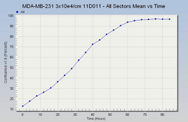
MDA-MB-231 cells are grown at 37◦C in Leibovitz’s L-15 medium supplemented with 2mM glutamine and 15% foetal bovine serum (FBS). This medium supports the growth of cells in environments without CO2 equilibration.
MDA-MB-231 cells should be seeded at a density between 1-3 x 10⁴ cells/cm2 and subcultured when 70-80% confluent.
Growth profile
|
Related cell lines |
ECACC catalogue number |
Description |
|---|---|---|
|
MDA-MB-157 |
Human breast medulla carcinoma |
|
|
MDA-MB-361 |
Human Caucasian breast adenocarcinoma |
Key references
-
CailleauR,Olive M, Cruciger QV. Long-term human breast carcinoma cell lines of metastatic origin: preliminary characterization. In Vitro, 1978. 14(11):911– 915.
-
Liu H, Zang C, Fenner MH, Possinger K, Elstner E. PPARgamma ligands and ATRA inhibit the invasion of human breast cancer cells in vitro. Breast Cancer Res Treat, 2003. 79(1):63-74.
-
Chavez KJ, Garimella SV,Lipkowitz Triple Negative Breast Cancer Cell Lines: One Tool in the Search for Better Treatment of Triple Negative Breast Cancer. Breast Dis, 2010. 32(1-2):35–48.
-
Holiday DL and Speirs V. Choosing the right cell line for breast cancer research. Breast Cancer Res, 2011. 13(14):215.
-
Harrell JC, Pfefferle AD,Zalles N,Prat A , Fan C, Khramtsov A, Olopade OI, Troester MA, Dudley AC, Perou CM. Endothelial-like properties of claudin-low breast cancer cells promote tumor vascular permeability and metastasis. Clin Exp Metastasis, 2014. 31(1):33–45.
-
Kenny PA, Lee GY, Myers CA, Neve RM, Semeiks JR, Spellman PT, Lorenz K, Lee EH, Barcellos-Hoff MH, Petersen OW, Gray JW, Bissell MJ. The morphologies of breast cancer cell lines in three-dimensional assays correlate with their profiles of gene expression. Mol Oncol, 2007. 1(1):84–96.
-
Simmons JK, Hildreth BE, Supsavhad W, Elshafae SM, Hassan BB, Dirksen WP, Toribio RE, Rosol TJ. Animal Models of Bone Metastasis. Vet Pathol, 2015. 52(5): 827-841.
-
Bos PD, Zhang XH, Nadal C, Shu W, Gomis RR, Nguyen DX, Minn AJ, van de Vijver MJ, Gerald WL, Foekens JA, Massagué J. Genes that mediate breast cancer metastatis to the brain. Nature, 2009. 459(7249):1005-1009.
-
Kang Y, Siegel PM, Shu W, Drobnjak M, Kakonen SM, Cordon-Cardo C, Guise TA, Massagué J. A multigenic program mediating breast cancer metastasis to bone. Cancer Cell, 2003. 3(6):537-549.
-
Minn AJ, Gupta GP, Siegel PM, Bos PD, Shu W, Giri DD, Viale A, Olshen AB, Gerald WL, Massagué J. Genes that mediate breast cancer metastasis to lung. Nature, 2005. 436(7050):518-524.
-
Minn AJ, Kang Y, Serganova I, Gupta GP, Giri DD, Doubrovin M, Ponomarev V, Gerald WL, Blasberg R, Massagué J. Distinct organ-specific metastatic potential of individual breast cancer cells and primary tumours. J Clin Invest, 2005. 115(1):44-55.